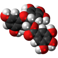
Epigallocatechin gallate (EGCG) is a is a polyphenol. It is a type of flavanol, specifically a type of catechin, the most abundant catechin found in tea. However, the compound is significantly degraded by steeping in boiling water, unlike related catechins.
Healing Properties
Digestion
Weight Loss
The combination of EGCG and Caffein suppresses body weight gain and fat accumulation.[1]
Anorexigenic (appetite reduction)
EGCG and caffeine has been shown to induce suppression of fat accumulation and a strong anorexigenic action. The anorexigenic effect may be brought about via inhibiting gastric motility.[1:1]
Disease / Symptom Treatment
Heart Attack
Reduces the Risk of Heart Attack
- The molecule EGCG has the ability to bind to proteins found in plaques linked to coronary artery disease and make them more soft and pliable making it easier for blood to flow through arteries and veins.[2]
Title: The combined administration of EGCG and caffeine induces not only suppression of fat accumulation but also anorexigenic action in mice
Author(s): Litong Liu, Kazutoshi Sayama
Institution(s): Graduate School of Science and Technology, Shizuoka University, 836 Ohya, Suruga-ku, Sizuoka 422-8529, Japan, College of Agriculture, Academic Institute, Shizuoka University, 836 Ohya, Suruga-ku, Shizuoka 422-8529, Japan
Publication: Journal of Functional Foods
Abstract: To elucidate the anorexigenic action and inhibitory effect of fat accumulation by epigallocatechin gallate (EGCG) and caffeine, including the optimal combination ratio and mechanism, fifteen diets with several concentrations of EGCG and/or caffeine were administered to mice for eight weeks. The 0.1% EGCG + 0.1% caffeine group showed the strongest suppression of food intake and a remarkable reduction of body weight and fat accumulation; therefore, the ratio was determined to be the optimal combination ratio. Moreover, serum glucagon-like peptide-1 (GLP-1) level and hypothalamic gene expression of pro-opiomelanocortin (POMC) were promoted by 0.1% EGCG + 0.1% caffeine. In conclusion, the combined treatment of 0.1% EGCG + 0.1% caffeine induces not only suppression of fat accumulation but also strong anorexigenic action in mice. The anorexigenic effect may be brought about via inhibiting gastric motility by GLP-1 and upregulating POMC in the hypothalamus.
Link: https://doi.org/10.1016/j.jff.2018.05.030
Citations: ↩︎ ↩︎Title: Epigallocatechin-3-gallate remodels apolipoprotein A-I amyloid fibrils into soluble oligomers in the presence of heparin
Author(s): David Townsend, Eleri Hughes, Geoffrey Akien, Katie L. Stewart, Sheena E. Radford, David Rochester, and David A. Middleton
Institutions: Lancaster University, United Kingdom, Emory University, United States, University of Leeds, United Kingdom, Chemistry, Lancaster University, United Kingdo
Publication: Journal of Biological Chemistry
Date: May 31, 2018
Abstract: Amyloid deposits of wild-type apolipoprotein A-I (apoA-I), the main protein component of high-density lipoprotein, accumulate in atherosclerotic plaques where they may contribute to coronary artery disease by increasing plaque burden and instability. Using CD analysis, solid-state NMR spectroscopy, and transmission EM, we report here a surprising cooperative effect of heparin and the green tea polyphenol (–)- epigallocatechin-3-gallate (EGCG), a known inhibitor and modulator of amyloid formation, on apoA-I fibrils. We found that heparin, a proxy for glycosaminoglycan (GAG) polysaccharides that co-localize ubiquitously with amyloid in vivo, accelerates the rate of apoA-I formation from monomeric protein and associates with insoluble fibrils. Mature, insoluble apoA-I fibrils bound EGCG (KD = 30 ± 3 μM; Bmax = 40 ± 3 μM), but EGCG did not alter the kinetics of apoA-I amyloid assembly from monomer in the presence or absence of heparin. EGCG selectively increased the mobility of specific backbone and side-chain sites of apoA-I fibrils formed in the absence of heparin, but the fibrils largely retained their original morphology and remained insoluble. By contrast, fibrils formed in the presence of heparin were mobilized extensively by the addition of equimolar EGCG, and the fibrils were remodeled into soluble 20-nm-diameter oligomers with a largely α-helical structure that were nontoxic to human umbilical artery endothelial cells. These results argue for a protective effect of EGCG on apoA-I amyloid associated with atherosclerosis and suggest that EGCG-induced remodeling of amyloid may be tightly regulated by GAGs and other amyloid co-factors in vivo, depending on EGCG bioavailability.
Link: http://dx.doi.org/10.1074/jbc.RA118.002038
Citations: ↩︎
