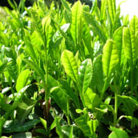
Green Tea (Camellia sinensis)
Healing Properties
Antioxidant
Green Tea contains antioxidants.[1]
Phenolics in green tea extract, including EGCG, ECG, EGC, EC, and gallic acid, may synergistically act on the antioxidant activity via chelating redox-active transition metal ions and scavenging reactive oxygen and nitrogen species.[2]
Skin Health
UV Protection
Green tea polyphenols were shown to reduce UV light-induced oxidative stress and immunosuppression.[1:1]
Topical treatment and oral consumption of green tea polyphenols (GTP) inhibits chemical carcinogen-or UV radiation-induced skin carcinogenesis.[1:2]
Anti-Obesity
Anorexigenic
Reduces appetite, resulting in lower food consumption, leading to weight loss.[3]
- EGCG & caffeine induces not only suppression of fat accumulation but also strong anorexigenic action.
- The anorexigenic effect may be brought about via inhibiting gastric motility.
- The combination of EGCG also suppresses body weight gain and fat accumulation.[3:1]
Brain Health
Acetylcholinesterase inhibitor (Anticholinesterase)
Green tea is known for its high content in tannins.
The tannins in green tea have been shown to greatly inhibit Acetylcholinesterase activity.[4]
- Acetylcholine is a chemical messenger in the brain and Acetylcholinesterase breaks down acetylcholine.
- Green Tea tannins inhibit the acetylcholinesterase enzyme from breaking down acetylcholine, thereby increases both the level and duration of action of the neurotransmitter acetylcholine.[4:1]
- There is a strong positive correlation between anticholinesterase activity and total condensed tannins.[4:2]
M2 Microglia Support
Green Tea with EGCG (Active Compound) Enhances the Expression of M2 phenotypes.[5]
Both EGCG and standardized green tea extract can increase CD206 expression, but standardized green tea extract outperforms EGCG.[5:1]
CD206 is considered a reliable marker for M2 activation in mice and humans because it is expressed mainly in M2 phenotypes.[5:2]
M2 phenotypes are activated by anti-inflammatory cytokines such as interleukin-4 (IL-4) and IL-13. These are also activated by T-helper 2 (Th2), which further upregulate scavenger receptors on M2 cell surfaces such as the mannose receptor (MRC1/CD206), and M2 phenotypes secrete anti-inflammatory cytokines such as resolution molecules.[5:3]
M2 microglia can inhibit inflammation, increase tissue repair and healing, and play a role in neurogenesis and functional repair. Recent studies have shown that M2 microglia assist neurogenesis in a post-stroke model; thus, so it is suitable for functional recovery.[5:4]
Neuroprotective
- Green tea polyphenols increased the activity of succinate dehydrogenase, as well as that of cytochrome c oxidase.[6]
- The cognitive deficit in animals which received green tea polyphenols was significantly lower than that in untreated animals.[6:1]
- A poison, sodium azide was introduced into the hippocampal tissue of animals, the development of cognitive deficits with a decrease in the activity of succinate dehydrogenase and cytochrome c oxidase is observed.[6:2]
- A course administration of green tea polyphenols increased the activity of succinate dehydrogenase and cytochrome-c-oxidase, which contributed to the restoration of cognitive abilities in animals.[6:3]
Cardioprotective
Green Tea Extract Improves Heart Muscle and May Help Treat Cardiomyopathies by Improving Mitochondrial Function.[7] [8]
Long-term in vivo administration of green tea catechin extract (GTE) resulted in an improvement of heart cell mechanical properties.
- This was measured by a significant increase in hyperdynamic cardiomyocyte contractility (an increase in the rate of shortening and re-lengthening of heart muscle cells, more specifically the fraction of shortening due to the amplitude of calcium transient, and the rate of cytosolic calcium removal).
Green Tea Extract supplementation has been shown to improve cardiomyocyte mechanics and intracellular calcium dynamics.
Cellular bioenergetics were found to be significantly improved:
- This study measured the maximal mitochondrial respiration rate and the cellular ATP level.
- The improvement of mitochondrial function was associated with increased levels of oxidative phosphorylation complexes.
- The cellular mitochondrial mass was unchanged.
- Mechanism of action: Green Tea Extract supplementation lowerered the expression of total phospholamban (PLB), which led to an increase of both the phosphorylated-PLB/PLB and the sarco-endoplasmic reticulum calcium ATPase/PLB ratios.
Green Tea Extract might be a valuable adjuvant tool for counteracting the occurrence and/or the progression of cardiomyopathies in which mitochondrial dysfunction and alteration of intracellular calcium dynamics constitute early pathogenic factors.
Endothelial Health
The molecule EGCG has the ability to bind to proteins found in plaques linked to coronary artery disease and make them more soft and pliable making it easier for blood to flow through arteries and veins.[4:3]
Skin Health
Green Tea’s Anti-skin aging activities are three-fold[2:1]
Green tea extract (composed mainly of epigallocatechin gallate, epigallocatechin, and epicatechin gallate) has shown positive activities against skin aging, including significant suppression of melanin production, potent antioxidant activities, and significant matrix metalloproteinase-2 (MMP2) inhibition (MMP2 is an enzyme involved in the breakdown of the extracellular collagen matrix). The study results have shown that green tea is a functional plant for utilisation as an anti-skin aging agent in the natural remedies, including food, health, and cosmetic products.[2:2]
Collagen Production
EGCG can suppress fibroblast proliferation and collagen production.[2:3]
Collagen is produced in the fibroblasts of the human dermis and is essential for healthy, firm skin.
Hyperpigmentation
Green tea may have potential as a pigment reducing agent with additional skin anti-aging properties.[2:4]
Hyperpigmentation in Caucasian and Asian skin is markedly associated with photoaging, a major type of extrinsic aging caused by exposure to UV irradiation.[2:5]
The phenolic constituents in green tea, including EGCG and gallic acid, have been shown to inhibit the pigment synthesis and tyrosinase expression.[2:6]
The decreased melanin production of green tea was mediated via the inhibitory effect of two melanogenic enzyme activities, tyrosinase and TRP-2, in the melanin biosynthesis pathway.[2:7]
Disease / Symptom Treatment
Diabetes
Green tea extract contains catechins which have anti-diabetic properties.[9]
Glucose Regulation
(Carbohydrate Digestion, Glucosidase inhibitor) The Catechins within Green Tea Extract exhibit a potent inhibition of α-glucosidase activity and moderate inhibition on α-amylase (these are glucosidases required for starch digestion). The overall effect of inhibition is to help reduce the flow of glucose from complex dietary carbohydrates into the bloodstream, diminishing the postprandial effect of starch consumption on blood glucose levels.[9:1]
Infections
Camellia sinensis extract shows high antibacterial activity against gram positive bacteria.[10]
Heart Disease
Helps reduce the Risk of Heart Attack.
Cardiomyapthy
Green Tea Extract Improves Heart Muscle and May Help Treat Cardiomyopathies by Improving Mitochondrial Function.[7:1] [8:1]
Long-term in vivo administration of green tea catechin extract (GTE) resulted in an improvement of heart cell mechanical properties.
- This was measured by a significant increase in hyperdynamic cardiomyocyte contractility (an increase in the rate of shortening and re-lengthening of heart muscle cells, more specifically the fraction of shortening due to the amplitude of calcium transient, and the rate of cytosolic calcium removal).
Green Tea Extract supplementation has been shown to improve cardiomyocyte mechanics and intracellular calcium dynamics.
Cellular bioenergetics were found to be significantly improved:
- This study measured the maximal mitochondrial respiration rate and the cellular ATP level.
- The improvement of mitochondrial function was associated with increased levels of oxidative phosphorylation complexes.
- The cellular mitochondrial mass was unchanged.
- Mechanism of action: Green Tea Extract supplementation lowerered the expression of total phospholamban (PLB), which led to an increase of both the phosphorylated-PLB/PLB and the sarco-endoplasmic reticulum calcium ATPase/PLB ratios.
Green Tea Extract might be a valuable adjuvant tool for counteracting the occurrence and/or the progression of cardiomyopathies in which mitochondrial dysfunction and alteration of intracellular calcium dynamics constitute early pathogenic factors.
Obesity
EGCG & caffeine induces suppression of fat accumulation.[3:2]
Skin Disease
Skin Cancer
In skin tumors, topical treament or oral consumption of green tea polyphenols or EGCG inhibits chemical carcinogen- or UV radiation-induced skin carcinogenesis.[5:5]
Keloids
Green tea polyphenol epigallocatechin-3-gallate (EGCG) suppresses keloid development without damaging normal skin.[5:6]
EGCG suppresses adhesion-related signaling in Normal Fibroblasts, without cytotoxic or proapoptotic activity.[5:7]
Green tea polyphenol epigallocatechin-3-gallate (EGCG) suppresses collagen production and proliferation in keloid fibroblasts via inhibition of the STAT3-signaling pathway.[5:8]
- Proliferation of Keloid Fibroblasts was more strongly suppressed by EGCG than was Normal Fibroblast proliferation.[5:9]
Keloid Fibroblasts proliferate more rapidly than Normal Fibroblasts. However, the enhanced proliferation of Keloid Fibroblasts was completely inhibited administration of EGCG.[5:10]
In addition, under low-density culture conditions, Keloid Fibroblasts proliferated more rapidly and generated larger colonies than Normal Fibroblasts, but the colony number of Keloid Fibroblasts was reduced by EGCG treatment.[5:11]
- EGCG more strongly inhibits proliferation of Keloid Fibroblasts than that of Normal Fibroblasts under in vitro culture conditions.[5:12]
EGCG prevents fibroblast outgrowth, collagen overproduction, and abnormal wound healing related to keloid pathology.[5:13]
EGCG suppresses the migratory potential of both normal and Keloid Fibroblasts, but this suppressive effect is greater in Keloid Fibroblasts.[5:14]
These findings indicate that EGCG efficiently suppresses wound healing-related events, such as collagen production, fibroblast outgrowth, and cell migration in Keloid Fibroblasts.[5:15]
Stroke
M2 Microglia Support
Recent studies have shown that M2 microglia assist neurogenesis in a post-stroke model; thus, so it is suitable for functional recovery.[5:16]
Green Tea with EGCG (Active Compound) Enhances the Expression of M2 phenotypes.[5:17]
Both EGCG and standardized green tea extract can increase CD206 expression, but standardized green tea extract outperforms EGCG.[5:18]
Title: An extensive Review of Sunscreen and Suntan Preparations
Publication: ARC Journal of Pharmaceutical Sciences (AJPS)
Date: April 2019
Study Type: Review
Author(s): AK Mohiuddin
Institution(s): World University of Bangladesh
Copy: archive, archive-mirror ↩︎ ↩︎ ↩︎Title: Green tea polyphenol epigallocatechin-3-gallate (EGCG) suppresses collagen production and proliferation in keloid fibroblasts via inhibition of the STAT3-signaling pathway
Publication: Journal of Investigative Dermatology
Date: October 2008
Study Type: Human Study: In Vitro, Animal Study: In Vitro
Author(s): Gyuman Park, Byung Sun Yoon, Jai-Hee Moon, Joo Young Noh, ChilHwan Oh, Seungkwon You
Institution(s): Korea University, Seoul, Korea; Chonnam National University, Kwangju, Korea; Gachon University of Medicine and Science, Incheon, Korea
Copy: archive, archive-mirror ↩︎ ↩︎ ↩︎ ↩︎ ↩︎ ↩︎ ↩︎ ↩︎Title: The combined administration of EGCG and caffeine induces not only suppression of fat accumulation but also anorexigenic action in mice
Publication: Journal of Functional Foods
Date: August, 2018
Study Type: Animal Study: In Vivo
Author(s): Litong Liu, Kazutoshi Sayama
Institution(s): Shizuoka University, Japan
Copy: archive, archive-mirror ↩︎ ↩︎ ↩︎Title: Epigallocatechin-3-gallate remodels apolipoprotein A-I amyloid fibrils into soluble oligomers in the presence of heparin
Publication: Journal of Biological Chemistry
Date: May 31, 2018
Study Type: Human Study: In Vitro
Author(s): David Townsend, Eleri Hughes, Geoffrey Akien, Katie L. Stewart, Sheena E. Radford, David Rochester, and David A. Middleton
Institutions: Lancaster University, United Kingdom; Emory University, United States; University of Leeds, United Kingdom
Copy: archive, archive-mirror ↩︎ ↩︎ ↩︎ ↩︎Title: The Effect of Green Tea with EGCG Active Compound in Enhancing the Expression of M2 Microglia Marker (CD206)
Publication: Neurology India: Publication of the Neurological Society of India
Date: May 2022
Study Type: Animal Study: In Vivo
Author(s): Abdulloh Machin, Dinda Divamillenia, Nurmawati Fatimah, Imam Susilo, D Agus Purwanto, Imam Subadi, Paulus Sugianto, Muhammad Hamdan, O Galuh Pratiwi, Dyah Fauziah, Kenia Izzawa
Institutions: Universitas Airlangga Hospital, Surabaya, Inonesia
Archive ↩︎ ↩︎ ↩︎ ↩︎ ↩︎ ↩︎ ↩︎ ↩︎ ↩︎ ↩︎ ↩︎ ↩︎ ↩︎ ↩︎ ↩︎ ↩︎ ↩︎ ↩︎ ↩︎Title: THE EFFECT OF GREEN TEA POLYPHENOLS ON THE CHANGE IN THE MITOCHONDRIAL FUNCTION OF HIPPOCAMPAL CELLS IN A DEFICIENCY IN THE ACTIVITY OF MITOCHONDRIAL COMPLEX IV
Publication: acta-medica-eurasica
Date: 2022
Study Type: Animal Study: In Vitro
Author(s): Pozdnyakov Dmitriy I.
Institutions: Department of Pharmacology, Pyatigorsk Medical and Pharmaceutical Institute, Russia
Copy: archive ↩︎ ↩︎ ↩︎ ↩︎Title: Long-Term Oral Administration of Theaphenon-E Improves Cardiomyocyte Mechanics and Calcium Dynamics by Affecting Phospholamban Phosphorylation and ATP Production
Publication: Karger: Cellular Physiology and Biochemistry
Date: July 2018
Study Type: Animal Study: In Vivo
Author(s): Bocchi L., Savi M., Naponelli V., Vilella R., Sgarbi G., Baracca A., Solaini G., Bettuzzi S., Rizzi F., Stilli D.
Institution(s): University of Parma, Parma, Italy; Fondazione Umberto Veronesi, Milan, Italy; University of Bologna, Italy; National Institute of Biostructure and Biosystems (INBB), Rome, Italy
Copy: archive, archive-mirror ↩︎ ↩︎Title: Effects of Standardized Green Tea Extract and Its Main Component, EGCG, on Mitochondrial Function and Contractile Performance of Healthy Rat Cardiomyocytes
Publication: MDPI: Nutrients
Date: July 2020
Study Type: Animal Study: In Vivo
Author(s): Rocchina Vilella, Gianluca Sgarbi, Valeria Naponelli, Monia Savi, Leonardo Bocchi, Francesca Liuzzi, Riccardo Righetti, Federico Quaini, Caterina Frati, Saverio Bettuzzi, Giancarlo Solaini, Donatella Stilli, Federica Rizzi, and Alessandra Baracca
Institution(s): University of Parma, Italy; University of Bologna, Italy; National Institute of Biostructure and Biosystems (INBB), Rome, Italy
Copy: archive, archive-mirror ↩︎ ↩︎Title: Grape Seed and Tea Extracts and Catechin 3-Gallates Are Potent Inhibitors of α-Amylase and α-Glucosidase Activity
Publication: American Chemical Society: Journal of Agricultural and Food Chemistry
Date: September 2012
Study Type: human Study: In Vitro
Author(s): Meltem Yilmazer-Musa, Anneke M. Griffith, Alexander J. Michels, Erik Schneider, and Balz Frei
Institution(s): Linus Pauling Institute, Oregon State University, Corvallis, Oregon, USA; USANA Health Sciences, Inc., Salt Lake City, Utah, USA
Copy: archive, archive-mirror ↩︎ ↩︎Title: Synergistic Antimicrobial Activity of Camellia sinensis and Juglans regia against Multidrug-Resistant Bacteria
Publication: PLOS ONE
Date: February 26, 2015
Study Type: Bacterial Study: In Vitro
Author(s): Amber Farooqui, Adnan Khan, Ilaria Borghetto, Shahana U. Kazmi, Salvatore Rubino, Bianca Paglietti
Institution(s): University of Sassari, Italy; King Faisal Specialist Hospital and Research Center, Riyadh, Saudi Arabia; University of Karachi, Pakistan; Shantou University Medical College, Guangdong, China
Copy: archive, archive-mirror ↩︎
